Cell cycle worksheet cells alive – Unveiling the intricacies of cellular reproduction, the Cell Cycle Worksheet: Cells Alive serves as an authoritative resource, delving into the fundamental processes that govern cell division. This comprehensive guide illuminates the various stages of the cell cycle, from interphase to cytokinesis, providing a profound understanding of the mechanisms that drive cell growth and proliferation.
As we journey through the intricacies of the cell cycle, we will explore the distinct phases of interphase, mitosis, and cytokinesis, unraveling the key events that orchestrate cell division. Furthermore, we will delve into the regulatory mechanisms that ensure the fidelity of cell cycle progression, examining the role of checkpoints, cyclins, and cyclin-dependent kinases.
Introduction to the Cell Cycle: Cell Cycle Worksheet Cells Alive
The cell cycle is a fundamental process by which cells grow, divide, and replace themselves. It is a continuous, orderly sequence of events that ensures the accurate duplication and distribution of genetic material to daughter cells.
The cell cycle consists of three main phases: interphase, mitosis, and cytokinesis. Interphase is the longest phase and is characterized by cell growth and DNA replication. Mitosis is the phase during which the duplicated chromosomes are separated and distributed to the daughter cells.
Cytokinesis is the final phase and involves the physical separation of the cytoplasm and organelles into two distinct cells.
Interphase
Interphase is divided into three sub-phases: G1, S, and G2. During the G1 phase, the cell grows and prepares for DNA replication. In the S phase, the DNA is replicated, resulting in two identical copies of each chromosome. The G2 phase is a period of further growth and preparation for mitosis.
Mitosis
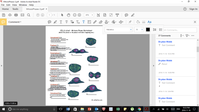
Mitosis is a complex process that involves the separation and distribution of duplicated chromosomes. It is divided into four distinct stages: prophase, metaphase, anaphase, and telophase. During prophase, the chromosomes condense and the nuclear envelope breaks down. In metaphase, the chromosomes align at the equator of the cell.
In anaphase, the sister chromatids of each chromosome separate and move to opposite poles of the cell. In telophase, the chromosomes reach the poles and the nuclear envelope reforms around each set of chromosomes.
Cytokinesis
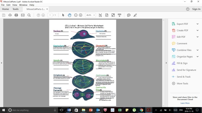
Cytokinesis is the final phase of the cell cycle and involves the physical separation of the cytoplasm and organelles into two distinct cells. In animal cells, cytokinesis occurs by a process called cleavage furrowing, where a contractile ring of actin and myosin filaments forms around the equator of the cell and pinches it in two.
In plant cells, cytokinesis occurs by the formation of a cell plate, which is a new cell wall that grows inward from the cell periphery.
Interphase
Interphase is the longest phase of the cell cycle, accounting for approximately 90% of the cell’s life. During interphase, the cell grows, replicates its DNA, and prepares for cell division.
Interphase is divided into three stages: G1, S, and G2.
G1 Phase
The G1 phase is the first stage of interphase. During G1, the cell grows and prepares for DNA replication. The cell synthesizes proteins and RNA, and it increases in size.
S Phase
The S phase is the second stage of interphase. During S phase, the cell replicates its DNA. Each chromosome is copied, resulting in two identical copies of each chromosome.
G2 Phase
The G2 phase is the third stage of interphase. During G2, the cell checks for DNA damage and repairs any errors. The cell also synthesizes proteins and RNA, and it increases in size.
Mitosis
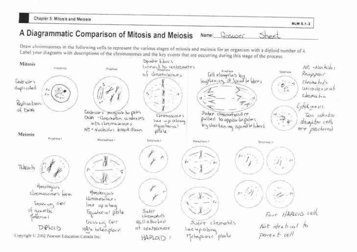
Mitosis is a type of cell division that results in two daughter cells that are genetically identical to the parent cell. It is a continuous process, but for the sake of study, it is divided into four distinct stages: prophase, metaphase, anaphase, and telophase.
Prophase, Cell cycle worksheet cells alive
Prophase is the longest and most complex stage of mitosis. During prophase, the chromatin in the nucleus condenses into visible chromosomes. The nuclear envelope breaks down, and the spindle fibers form. The spindle fibers are made of microtubules and are responsible for separating the chromosomes during anaphase.
Metaphase
During metaphase, the chromosomes line up in the center of the cell. The spindle fibers attach to the chromosomes at their centromeres. The centromeres are the regions of the chromosomes where the sister chromatids are joined together.
Anaphase
During anaphase, the sister chromatids separate and move to opposite poles of the cell. The spindle fibers shorten, pulling the chromosomes apart.
Telophase
During telophase, the chromosomes reach the poles of the cell. The spindle fibers disappear, and the nuclear envelope reforms around each set of chromosomes. The chromosomes then begin to decondense, and the cell membrane pinches in the middle, dividing the cell into two daughter cells.
Cytokinesis
Cytokinesis is the physical separation of the cytoplasm and organelles into two distinct daughter cells, following mitosis or meiosis.
In animal cells, cytokinesis occurs by a process called furrow formation. A contractile ring of microfilaments forms just inside the plasma membrane and constricts, pinching the cell in two. In plant cells, cytokinesis occurs by cell plate formation. Vesicles containing cell wall material accumulate at the middle of the cell and fuse to form a new cell wall, dividing the cell into two.
Role of the Cell Plate and Contractile Ring in Cytokinesis
The cell plateis a structure that forms between the two daughter cells during cytokinesis in plant cells. It is made up of vesicles containing cell wall material, which fuse together to form a new cell wall. The contractile ring is a structure that forms just inside the plasma membrane of animal cells during cytokinesis.
It is made up of microfilaments, which contract to pinch the cell in two.
Regulation of the Cell Cycle
The cell cycle is tightly regulated to ensure that cells divide only when they are healthy and have the necessary resources. Several checkpoints are located throughout the cell cycle to monitor cell growth, DNA replication, and DNA damage.
Key Checkpoints in the Cell Cycle
The key checkpoints in the cell cycle are:
- G1 Checkpoint:Occurs at the end of G1 phase. It checks for cell growth and DNA damage before allowing the cell to enter S phase.
- S Checkpoint:Occurs during S phase. It checks for DNA replication errors before allowing the cell to enter G2 phase.
- G2/M Checkpoint:Occurs at the end of G2 phase. It checks for DNA damage and ensures that the cell is ready to enter mitosis.
- M Checkpoint:Occurs during mitosis. It checks for errors in chromosome segregation before allowing the cell to enter cytokinesis.
If any of these checkpoints detect a problem, the cell cycle will be arrested until the problem is resolved.
Role of Cyclins and Cyclin-Dependent Kinases (CDKs)
Cyclins and cyclin-dependent kinases (CDKs) are key regulators of the cell cycle. Cyclins are proteins whose levels fluctuate throughout the cell cycle. CDKs are enzymes that phosphorylate other proteins, which can activate or deactivate them.
Cyclins and CDKs form complexes that are active at specific stages of the cell cycle. These complexes phosphorylate target proteins, which in turn regulate the progression of the cell cycle. For example, the cyclin-CDK complex that is active in S phase phosphorylates proteins that are involved in DNA replication.
The activity of cyclin-CDK complexes is also regulated by other proteins, such as tumor suppressor proteins and oncogenes. Tumor suppressor proteins can inhibit the activity of cyclin-CDK complexes, while oncogenes can activate them.
The regulation of the cell cycle by cyclins and CDKs is essential for ensuring that cells divide only when they are healthy and have the necessary resources.
Applications of Cell Cycle Analysis
Cell cycle analysis is a powerful tool in biomedical research and clinical practice. It provides valuable insights into the health and behavior of cells, enabling researchers and clinicians to understand and diagnose a wide range of conditions.Cell cycle analysis can be used to diagnose and monitor diseases such as cancer, autoimmune disorders, and infectious diseases.
By examining the cell cycle distribution of a patient’s cells, clinicians can identify abnormalities that may indicate disease. For example, in cancer, an increased proportion of cells in the S phase or G2/M phase may indicate rapid cell proliferation and uncontrolled growth.
Helpful Answers
What is the significance of the cell cycle?
The cell cycle is essential for maintaining tissue homeostasis, ensuring the proper growth and development of multicellular organisms. It allows cells to divide and produce daughter cells, replenishing old or damaged cells and facilitating tissue repair.
How is the cell cycle regulated?
The cell cycle is tightly regulated by a complex network of checkpoints, cyclins, and cyclin-dependent kinases (CDKs). These regulatory mechanisms ensure that the cell cycle proceeds in an orderly and controlled manner, preventing errors that could lead to genomic instability or uncontrolled cell growth.
What are the applications of cell cycle analysis?
Cell cycle analysis plays a crucial role in biomedical research and clinical practice. It is used to diagnose and monitor diseases such as cancer, where abnormal cell cycle progression can contribute to uncontrolled cell proliferation and tumor formation.
